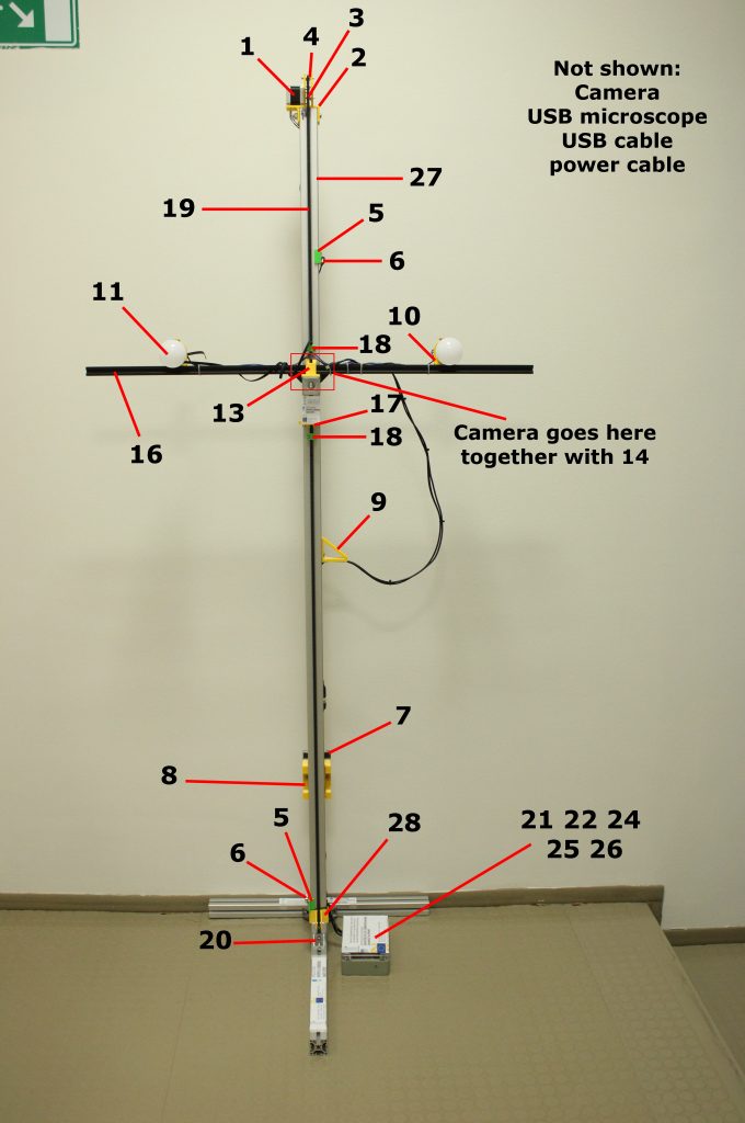
In the past days, our team has been subjecting the hardware and software that has been developed for our project to rigorous testing. Multiple bugs have been fixed and the dermoscope stand has been modified so that the 3D-printed components have been reinforced and all cables and plugs now join in a box at the stand’s base.
The videos below show the process of capturing an image from one perspective. The camera moves with a speed of about 10 cm/s, meaning the patient’s whole-body scan (from four angles – the front, the back and both sides) takes about 2 minutes.
The following video shows the graphic user interface (GUI) which guides the user through capturing a whole-body scan.
Instructions and parts
The open-source computer program which was developed is freely available on the online platform GitLab (link, zip). It enables the user to capture an image of the whole body, after which they can annotate individual skin lesions and capture an additional, magnified image with a USB microscope. After capturing the images, the user can add comments and compare individual exams in order to determine which skin changes require further treatment or investigation.
The list of components we used is accessible here. We hope it serves as a guide for anyone wishing to construct their own dermoscope stand or simply as a starting point for future projects.
